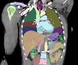
 CTACardio / actual labels
Image → anatomy
A navigable anatomy tree, three linked image planes and matching 3D surfaces. Start with real CT and MRI examples.
Explore the imaging atlas ↗
Liver · gallbladder · native vessels
Loading…
From medical images to anatomical assets to the mechanics of living tissue. A connected library for people building what comes next.
Keep the scan, the anatomical structure and the physical model connected — with their sources and assumptions intact.
CTACardio / actual labelsA navigable anatomy tree, three linked image planes and matching 3D surfaces. Start with real CT and MRI examples.
Explore the imaging atlas ↗Reusable geometry with units, materials, semantic labels and provenance. Physical models carry explicit constitutive assumptions.
Inside an asset ↗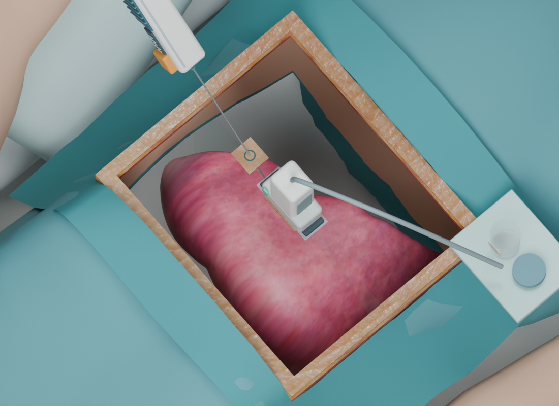 APPLICATION CASE / LIVER BIOPSY
Explore a concrete procedure: patient, target lesion, ultrasound probe and biopsy needle. Follow the approach, tissue sampling and withdrawal.
Explore the biopsy case ↗Attach a physical meaning to an anatomical structure: constitutive law, tissue parameters, contact, support and solver evidence. Know exactly what each model represents.
Explore biopsy, embolization and surgery through the anatomy, instruments and sequence that define each application.
Choose an example. Follow a structure. Watch a solved experiment.